Quantitative and Microscopic Imaging Center
Many of today's biomedical problems are best addressed, and sometimes can only be addressed, using the interdisciplinary approaches. This research center is based on the Xia Research Lab in the Physics Department at Oakland University. We emphasize the correlations among physics, chemistry, mechanical engineering, biology and medicine, using an interdisciplinary approach, including:
- Microscopic MRI (µMRI) - It is totally non-invasive and non-destructive, and shares the same physics principles and engineering architectures with the clinical whole-body MRI in hospitals. The 2D resolution of this system can be as fine as 10 µm.
- Polarized light microscopy (PLM) - It is a gold standard in histology, with optical resolution to visualize the morphology of the fibril structures in cartilage.
- Fourier-transform infrared imaging (FTIRI) - It is a chemical imaging technique, which is capable of detecting the earliest degradation before any morphological or clinical lesion.
- Microscopic computer tomography (µCT) - It shares the same physics principles and engineering architectures with the clinical whole-body CT in hospitals, which permits the imaging of both cartilage and bone.
- Mechanical imaging system - It is a home-built imaging system for bio-mechanical properties, which is capable of imaging the depth-dependent compressive modulus in connective tissues.
These state-of-the-art research instruments are sitting next to each other in the lab (the first floor of Hannah Hall). We know the precise protocols in each technique. We choose the best combination among these technical capabilities to research a particular problem. We aim to provide answers that are meaningful and without ambiguity. Please contact the Director of the QMIC, Prof Yang Xia ([email protected]), to arrange for a visit to the Facility, and to discuss your project, any user fee, and any available schedule.
We have a Bruker AVANCE IIIHD Console, a 7Tesla-89mm magnet and two sets of micro-imaging gradient coils. The system runs ParaVision 6.
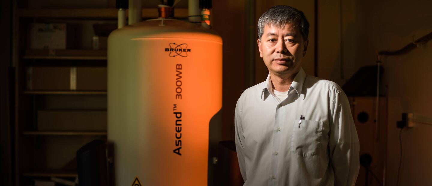
The lab started with a used Bruker AMX console and an Oxford magnet when Dr. Xia joined Oakland University in 1994. The lab kept the ancient AMX working until 2007, which was upgraded to the AVANCE II console. In May 2011, the equipment was replaced with a Bruker 7T/89mm Ascend 300WB (the series number on our Ascend magnet is #1). In 2018, the lab upgraded the AVANCE II console to AVANCE IIIHD.
A number of one-of-a-kind devices have been designed and built in our µMRI lab, to image tissue specimens under loading or anisotropically. A number of imaging and spectroscopy pulse sequences have also been purposely developed and extensively employed in the lab, to carry out the T1, T1Ï, T2, diffusion, and double-quantum-filtered experiments.
The lab has a Leica DM polarized light microscope, which has two digital imaging systems: (a) a home-built system with a thermoelectrically cooled Sensys camera, where we write our own software to analyze the binary images; and (b) a commercial Abrio system from Cambridge Research & Instrumentation (Wobum, MA), on which we can analyze the proprietary data from CRI.
For each tissue section, our protocol calculates two quantitative images: the angle (in degree) and the retardation (in nm) of the tissue section. For articular cartilage, the 2D angle map displays the orientation of the collagen fibrils in cartilage; and the 2D retardation map illustrates the organization of the fibril and macromolecules in cartilage. We routinely use magnification between 0.25µm/pixel (sufficient for quantitative mapping of the chondrocytes/territorial matrix in the tissue) and 2µm/pixel (suitable for whole-thickness morphology).
The lab has a Skyscan 1174 system, which has a 20-50kV x-ray source and 1.3MB CCD camera. The spatial resolution is 6-30 µm isotropic. The sample size can be 5-30mm in diameter and max 50 mm in length. Several 2D and 3D data analyzing software are available for this imaging system.
In 2015, we published a journal paper on the quantification of GAG molecules in cartilage using µCT, which for the first time quantified the depth-dependent GAG concentration and investigated the zonal variations in both healthy and degraded cartilage.
This GAG quantification project by µCT will facilitate future multi-disciplinary investigations in the degradation of in vivo, ex vivo and in situ cartilage and bone systems.
We have a PerkinElmer Spotlight 300 imager, which consists of a classical FTIR spectrophotometer coupled to an infrared microscope. The pixel size of the liquid-nitrogen-cooled detector is 6.25 µm. We also have an ATR attachment, which can improve the pixel resolution in this system down to 1.56µm.
We have published 16 journal papers on the studies of cartilage and its degradation by FTIRI.
This system is built around an ancient Nikon inverted microscope. Two cameras are connected with the microscope: a Spot Insight Monochrome camera from Diagnostic Instrument (14 bit AD with 20 MHz capture), and a Canon digital Rebel (for movies and large color images).
The images from the Spot camera are captured by the computer on the left of the microscope. On the right side of the microscope, the computer controls a microscopic indenter, which can compress cartilage either in discrete steps or continuously. The final devices on the right control a load sensor.
Our Biomechanics Testing Lab has an EnduraTec ELF3200 testing system, which has 0.02N force resolution and 0.5µm displacement resolution. This system is capable of both static and dynamic testing.
This system itself does not have any imaging capability. However, if you use a small enough indenter and move the sample slowly along a certain trajectory, you are doing localized measurement - you are doing a (low-resolution) imaging! Our smallest indenter is about 60µm in diameter.
The lab has the full sets of equipment for histological sectioning (paraffin section, frozen section, thick-section) and histochemical analysis.
The major items in histology include a tissue-infiltration processor (Sakura VIP E Tissue-TEC), a paraffin-embedding center (Sakura TEC 5), a paraffin-tissue microtome (Microm HM 325), a cryostat (Leica CM1950), and a vibrating blade microtome (Leica VT1000S).
The major items for histochemistry include a UV-VIS Spectrophotometer (Shimadzu 1650 PC) and two microbalances. Other major items in this lab include a low-magnification stereo optical microscope (Olympus), a cell culture incubator (Queue), a gravity convection oven (Blue M), a water bath (SP), an analytical balance (Mettler), an ultrasonic cleaner, and a -80˚C freezer (Harris).
We have established a number of biochemical and histochemical protocols in the lab, to measure the concentrations of PG, collagen, and water in cartilage. We have access to several (4˚C) cold-rooms that are located in the adjacent buildings, to carry out the measurement of water content in tissue blocks.
In my µMRI Lab, we also have a double-door freeze-refrigerator (Revco flammable storage) and a -80˚C freezer (Sanyo) for sample storage, and various machine and hand tools (including a low-speed MTI diamond saw and a low-speed grinder) for specimen preparation.
Original Gadgets
We love to build little gadgets for our experiments. These gadgets often contain both mechanical parts (serving a specific purpose, such as in situ loading or rotation) as well as electronics parts (an extension of the imaging probe or rf coil). The College of Arts and Sciences at Oakland University has a well-equipped Machine Workshop that is located just opposite our NMR lab.
We have a 500MHz Network/Spectrum/Impedance Analyzer (Agilent 4395A), a 400 MHz oscilloscope (Tektronic TDS 380), and many other relevant devices.
We have tens of computers and workstations in the labs running different operating systems (Mac OS, Windows, Unix, Linux). The computers in the µMRI Lab are isolated by a set of secure firewalls, which is then connected to the campus Ethernet.
Shared Equipment
We have been using a Perkin Elmer Optima 7000DV Inductively Coupled Plasma - Optical Emission Spectrometer, that belongs to the Department of Chemistry at OU. We have published several journal papers that documented the use of data from this instrument (e.g., Phys. Med. Biol. 58 (2013) 4535–4547).
We can request access to several electron, light and confocal fluorescence microscopes at the Eye Research Institute and Biology Department at OU. We have been using these microscopes for the live/dead cell imaging in cartilage.
The Department of Chemistry at OU has two multinuclear NMR spectroscopic systems, a Bruker AVANCE 200MHz system and a Bruker AVANCE III 400MHz system. Each system has a narrow-bore magnet. We can request time on these systems for any advanced spectroscopy experiment.
We have access to several whole-body human MRI scanners at the local hospitals (Henry Ford Hospital in Detroit, Wayne State University Medical School MRI Center in Detroit, William Beaumont Hospital in Royal Oak). There are also two 7T animal scanners at the two hospitals.
Department of Physics
146 Library Drive
Rochester, MI 48309
(location map)
(248) 370-3416
Fax: (248) 370-3408
[email protected]
Department Chair:
Professor Andrei Slavin
Society of Physics Students:
Office: 288 Hannah Hall (HH)


